Discussion #4989
Knee Anatomy & Biomechanics
Description
About This Video
Transcript
Read Full Transcript
Hello, my name is Dr. Brent Anderson, and I am excited to be with you on "Pilates Anytime" talking about the knee. And I hope to take you on a journey from understanding the knee in its normal anatomy and its development from childhood to old age, as well as the injuries that we incur and the interventions or treatments there. One of the key things that I like to bring to "Pilates Anytime" and to my clients, my patients is the idea that education is power. When you are educated about your knee, how your knee normally works, what is that disease, What is that surgery, what does rehabilitation include, why pain, what is pain for, and how does pain relate to this, that's usually what our concern is, it's usually what brings us to the doctor. So this presentation, I want to be at about a medium level of science.
It is an education for you as an individual about your own knee, about your own health and your knee, but also about your clients because a lot of you are Pilate's teachers and physical therapists and occupational therapists that are gonna be listening to this workshop. So I hope to take you on a great journey and we are gonna get started with anatomy. We often refer to this as the kinesiology of the knee, having to do with the science, ology, of movement, kinase, as it pertains to the knee. So I always like to break the word apart a little bit in its Latin form, to understand that we're gonna be studying the science of movement as it pertains to the knee. And to do that, we have to understand the anatomy as well as the biomechanics.
And when we talk about biomechanics, we break it into two particular categories, what we refer to as osteokinematics, meaning the movement of the bones, as well as arthrokinematics, which is the movement of the joint. I think it's very important for us to distinguish this and I'll go into more details. And we're also going to relate it to a lay person's term that helps us in teaching and understanding it that we refer to as bone rhythms. The anatomy of the knee is quite complex, actually. It's one of the most complex joints in our body.
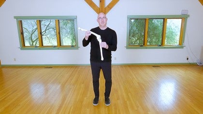
It has two condyles that are on the femur, the femur is the long bone of the thigh, and those two condyles articulate with the tibia. So when we look at the femur, the femur, as I mentioned, has two condyles in the bottom of it. Here appears is the head of the femur that is part of the hip bone that you're familiar with, if you watched my other lecture on "So You're Thinking About a Hip Replacement," also on "Pilates Anytime," and these two condyles are particularly designed to deal with actually a very complex biomechanics of the knee. We're gonna try to keep it simple but just understanding that they're two rounded surfaces that are going to articulate with the tibia and the tibia has two hollow surfaces on the top of it, and these surfaces come together, and this is what makes up the knee joint. So the tibia moving on the femur or the femur moving on the tibia are the primary knee joint, and it can move in either direction.
We can look at something that's open, we call this an open chain. I'll talk about a little bit more in the few slides. We also can talk about a closed chain where the foot is on the ground, and now the femur is moving on top of that joint. Now, what holds these bones together? Well, when we look at the ligaments or the capsule, they actually are designed very specifically to connect the femur to the tibia, and we're gonna talk about four ligaments in particular.
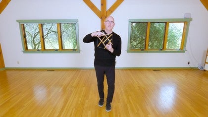
So on the slides, we can see the ligaments in the first picture on your left and we notice that there are ligaments on each side of the joint, and again, connecting the femur to the tibia, and those are called the medial collateral on the inside of the knee, so that would be on the inside of our knees connecting the femur to the tibia, and we have the lateral collateral ligament, again, collateral means on the sides, the lateral collateral beyond the lateral side of the knee joint connecting again the femur to the tibia, and they protect the knee from forces in the coronal plane. So if I was playing soccer and somebody ran into me on the side, my medial collateral ligament protects the knee from collapsing towards the center of my body. If I was falling downstairs and I went to the side this way, the lateral collateral ligament would protect my knee from getting damaged going towards the outside but the movement always in the coronal plane. The other two ligaments are the cruciate ligaments. The cruciate means crossing, are the two ligaments in the middle of the knee, and you have an anterior cruciate, anterior in front, posterior cruciate in back posterior, and they cross over and they protect anterior and posterior migration.
So that's more when, if I got hit from behind playing American football or soccer or football, that that movement of pushing my tibia forward on my knee is protected and checked by the anterior cruciate ligament. If I was in a car sitting in the front seat and I had my shins up against the dashboard, and I got an accident and the car pushed my tibias back towards my knee, that is the posterior cruciate ligament, a little more rare injury, that is protecting me from that tibia being displaced. And so the ligaments' job are to be relatively rigid and that rigidity protects the end of range of motion in our knees, so they're there to protect us. And then around the knee, or any joint, is what we refer to as a capsule, and the capsule is what protects the joint from movement as well, that we don't want. It also has a lining inside of it, the synovial lining that provides lubrication.
If we go down a little bit deeper in the anatomy, we see that there's a little disc on top of the tibia, and that little disc on top of the tibia on both side is referred to as the meniscus, and we have a medial and lateral meniscus of each knee. You often hear of people referring to meniscal tears, and we'll talk about that when we go into the pathology section, but the meniscus is a disc. It's hydrated and it has blood in certain parts of it, in other parts, it doesn't. So if you think of the labrum in the hip, the labrum in the shoulder, the disc in your spine, the disc in your jaw, these are all similar materials of our body that provide shock absorption. They enhance stability because they make a deeper bowl for the condyles to sit in, so it minimizes translation and movement of the knee, and I'll make sense out of this later as we talk that if I have my meniscus removed, then the likelihood of my condyles moving excessively on top of the tibia are greatly increased and that could lead to something like degenerative arthritis, and we'll talk about that later, but again, this idea of why do we have these structures, why do they exist inside of our knee.
Another really important part of anatomy of the knee is the patella, the kneecap, and the patella is strong over the joint with ligaments or tendons that connect to the quadriceps and to the tibial tuberosity of the knee. And you can see in this picture on the right, you can see how the patella sits on top of the groove of the femur. So you hear people sometimes saying, my patella dislocated, or my patella sublux, or I have patella femoral pain, or I have chondromalacia of my patella, this is talking about the relationship between the patella bone and the notch inside the femur that you see on this picture. So a lot of times when we lose alignment or we don't have good strength in our hips, that will cause a sort of an internal rotation of the femur and that makes that patella look like a slingshot and creates a tension in there that often could be very uncomfortable or painful, so we're gonna talk more about those pathologies as we understand them, so the patella is a pulley function. What we see is that it creates a lever and makes the lever come away from the center of the knee joint, so if here's my knee joint, here's my patella, it gives me power.
When the quadriceps pull on that patella, it's much more powerful to lift up the foot or the leg if I was going to kick or if I'm going to squat, it increases the fulcrum so that I have more power. So when people have their patella removed, they'll end up having less power, even that one inch of distance from the center of the joint can make huge differences in the amount of power in the leg. So when the patella is injured, or the tendon is injured and it's giving messages of pain, you can imagine how you would also see weakness associated with that pain. Another really important part of the anatomy in the knee is the fibula. The fibula is parallel to the tibia and it has the lateral collateral ligament comes down attaches to it as well, and so do the muscles.
When we look at the muscles of the knee, we typically are talking about the extensor muscles, which are gonna be the quadriceps, the sartorius muscle in front, the rectus femoris, those are all going to straighten the knee, or if I'm squatting, going to help me straighten my knee in squatting. We have the adductor muscles, so the gracilis and the brevis and the longest muscle, that help us with adduction and it's gracilis and the longest that cross over the knee, right, so those two muscles of the adductors provide that strength. And then we also have the IT band on the side, with the tensor fasciae latae, and that tendon comes all the way down and crosses over the knee joint as well and attaches into that fibular head. And then lastly, we have the hamstrings or the knee flexors that we have coming down and helping us to flex the knee and those also on the lateral side attached to the fibula. So one of the things that I like to bring people's attention to, especially if you're a therapist or a seasoned Pilates teacher, is that that fibula often can create lateral knee pain that is often misinterpreted as an internal derangement of the knee.
I've seen people get surgeries unnecessarily and not had, didn't need it and just dealing with a little bit of mobility and some pointing and flexing of the ankle might be enough to really reduce those symptoms and avoid surgery altogether. So we'll talk about that when we get to working with the fibula. And that pretty much covers the anatomy of the knee for the level that we're gonna be talking about it. I do wanna, there's one part that I forgot and that is that the gastrocnemius muscles, we often think of them hooking to the Achilles tendon and helping us with plantar flexion of the ankles or heel raises, that also crosses over the back of the knee and attaches on to the condyles of the femur on the backside of it or the posterior side. And so those are all the muscles that we're gonna primarily be working with as it pertains to knee health.
Osteokinematics. I mentioned that when we look at the Latin word, osteo means bone, kinematics means movement, so it is movement of the bone. When we think of the active range of motion of the knee, we often think of flexion and extension. It is a joint that moves in the sagittal plane. So if I'm kicking something, if I'm squatting, if I'm walking, or running, all of those movements of the knee are in the sagittal plane.
We also have a little bit of rotation that could also be considered an osteokinematic relationship, and the way that I remember these is I'm thinking of what are the bones moving through space. So if I bend my knee, I am flexing the knee, that is an osteo kinematic movement, if I straighten the leg or the tibia out on the femur, that is extension of the leg. And those are the two primary movements that we refer to in our knees. The knee is also sort of similar to the elbow. We think of the same thing, we flex and extend the elbows, that's the movement that's available in those joints.
So from an osteokinematic standpoint, knee osteokinematics consist of flexion and extension of the knee and the lower extremity. Before we go into the arthrokinematics, I want to talk about the kinetic chain, and you'll often hear people talking about, it's an open kinetic chain, it's a closed kinetic chain, it's a pseudo-closed chain. These are terms that we use to define where the movement occurs and where the resistance or the force is coming from. So when we look at things like an open-chain activity, what we're saying is that the distal part of that extremity is moving through space. So if I'm throwing a ball, if I'm kicking a ball, a lot of other activities involve that open chain movement.
It's the more complex. If I'm feeding myself, if I'm throwing things, moving my hands, those are all considered open-chain activities, open kinetic chain activities. And so, as it pertains to the knee, it's important for us to understand that walking, the major portion of walking is the swing phase and that is considered open chain, when the leg is swinging through. When the leg that's weight bearing in the ground forces, is considered a closed chain. So by definition, closed-chain kinetics is when the distal part of the extremity is fixed and the proximal or closer to the trunk is moving on the distal.
So for example, in the lower extremity, a squat is 100% closed-chain activity. If I was to do a pushup on the floor with my hands fixed on the floor and my body doing a push up, that would be considered a closed-chain activity. And so in walking and running, we have a mixture of those with our lower extremities. When we're in a swing phase of gait, that legs coming through and moving, as long as it's in the air, it's in an open chain, as soon as it hits the ground, now it's closed chain. So what we notice is that as we move faster, for example, going from walking to running, we will have more open chain than closed-chain activity in the lower extremity.
And it's important to understand these terms because in Pilates, we create closed and open-chain activities. We also create a new one that we refer to as pseudo, P-S-E-U-D-O, pseudo-closed chain or open chain. So for example, on the reformer, I'll let you think about this for a second, on the reformer doing footwork, is that a closed, open, or pseudo-closed chain for the lower extremity? Think about the definition, the distal part of the extremity or the furthest part away would be the foot, is fixed on a surface that is not moving, and the proximal part of the body is moving on the fixed portion. So if you thought footwork on the reformer is a closed-chain activity for the lower extremity, you are right.
Let's think about another one. Let's think about on the chair doing a single leg press. Our foot now is on the pedal, the pedal is a fixed arc in space, that fixed arc in space has resistance from the springs, and we can vary that, but the leg moving through that space, is that going to be an open-chain movement where it's completely free to move through space, is it a closed chain where the distal extremity is fixed, or is it something in between that we might call a pseudo chain activity? And if you thought pseudo-closed chain, you thought right. Another pseudo-closed chain activity could be something as simple as riding a bike.
It has a fixed arc with the pedals, the feet are fixed on the pedals, it has to go through that arc of space, and it's neither closed nor open, and so we call it a pseudo-closed chain. And I'll be using those terms as we go through and talk about bone rhythms and as we go through and talk about the exercises and why we selected them. But let me give you a little more information about why we differentiate between closed and open-chain activities. For example, did you know that closed-chain activities provides a lot more proprioception or awareness to the joint than open-chain activities? So if I was just starting my rehabilitation after an injury or a surgery, it might make sense to start with a closed-chain activity, closed chain because it's gonna give me a lot more awareness into my central nervous system as to where my body is in space.
I remember the first time that I had an orthopedic surgery, I remember thinking, asking myself, "Whose leg is that? It's not my leg. I don't feel it, I don't recognize it," and that is a common experience after a knee surgery or an ankle surgery and we need to be able to rebuild that awareness and that proprioception, we call it. Proprioception means awareness of self, right, so awareness of ourself internally so that we can rehabilitate and return to function. I hope that makes sense with the kinetic chain activities 'cause this really helps us in our decision as to what we're gonna do. Now, another example of the kinetic chain in Pilates is open chain.
So for example, if I had my feet in straps, the feet in straps have a fixed arc, so they could be a pseudo closed, pseudo open chain, but what would happen if I put my feet in the long springs on the trapeze table and I was able to move them in any direction, or if I used the connector cords or cables on the reformer and they allow me to have independence in where my leg goes relative to the other leg, that starts to become more and more open chain and more and more challenging, more and more difficult for the individual to be able to do that. Now that said, the majority of our activities, believe it or not, are open chain. So even though in acute rehabilitation, early rehabilitation we might wanna start with closed-chain activities, we eventually have to progress our clients or ourselves into open-chain functional activities that we would see while we're walking, running, jumping, going upstairs, downstairs, playing sports. All of those have that mixture of closed and open-chain activities. It's time to dive into arthrokinematics, and I love arthrokinematics.
This is the area that is the small differences make big changes. I wanna start off with a story. When once was working with a very elite athlete, a professional basketball player who had multiple knee surgeries and the last of which was a very successful surgery and yet the patient complained that he still didn't have what he called his blow, like being able to jump up and get his arm over the rim like he used to. And as I was working with him, I had him on the chair and I had him with an assisted lounge, so his affected leg was up on top of the chair, his unaffected leg was on the pedal, I had all the springs on, he was six foot eight, a big guy, and all I did was play a little bit with the spiral in the glide in the knee that had just had the surgery about four months before and something clicked in him that everything turned on. And the next day, he went to practice and he said that everything came back and he was explosive and able to play ball and he played one more year before he retired and was very happy about that.
I think about that often because sometimes there are these little things, when we talk about arthrokinematics or little things, that can make the big difference so let's jump in. Today, talking about arthrokinematics of the knee, we're primarily talking about three motions. Sometimes we use different names for them but I'm gonna be using spiral, I'm gonna be using glide, and roll. Sometimes, we also use the word spin for roll. So when we look at the knee, the knee has a very unique anatomy because the femur, and if you think about the human anatomy, the head of the femur has the neck and it takes the greater trochanter out to the side.
That's the bone we feel out here and some people refer to as their hips. The hip joint actually is very close inside. But because of that angle and we're bipedal animals, the medial condyle is much bigger than the lateral condyle, that allows us to shear up the change into the tibia and for us to stand straight, and the way we can test that is simply by just lifting one foot off at a time and we have very little shifting. A monkey has to be weigh out here to be able to shift their weight 'cause their pelvis in their center gravity is much wider, right? So we have this very unique condyle, that medial condyle, I think chickens do too, and that allows for what we call, or the first arthrokinematic which is spiral.
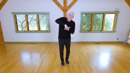
So when the knee bends, here we are bending, I'm bending down, the femur spirals laterally, the tibia responds by spiraling medially. When I straighten up, the femur spirals medially and the tibia spirals laterally until it's back up on top of each other. So let's rehearse that, stand up with me and just play this game. When we squat down, our arms are gonna represent our femurs, and I'm squatting down, the femur is spiraling laterally, when I straighten up, the femur spirals medially. When I squat, femur spirals laterally, and I come up, femur spirals medially.
Now in all joints of the body, if one bone is moving in one direction, in order for the joint to work properly, the other bone has to spiral or move in the other direction. So as I said, if the femur is spiraling laterally, the tibia is spiraling medially, when I stand up, the tibia spirals laterally and the femur spirals medially. When I use these words spiral, sometimes it gets confusing and people think we're talking about osteokinematic motion of external and internal rotation of the hip, and we're not, we're talking about something that happens inside the joint for it to bend successfully. So what that's gonna look like again is I'm not turning the whole leg out, it's something that happens right in the joint, and the way we know it's working is because we maintain that dynamic alignment of hip over knee, over ankle, over second toe. And that doesn't matter if I'm in turnout position or even if I'm turned in, I still can maintain that dynamic alignment.
So that's number one, is spiral. Number two is glide. So when I flex my knee, the tibia glides forward on the tibia, and if it's gonna glide forward on the tibia, that's the same thing as the tibia gliding posteriorly, otherwise there's no movement, and when I straighten, tibia glides forward, femur glides posteriorly to get back on top of it. So the second accessory motion of the knee is glide, right? As I bend, the femurs gonna glide forward, tibia glides back, as I straighten, it's gonna glide back into that starting position.
That's number two. Number three is a roll or a spin. So we said, these are like wheels on the condyles, these are like the treads when we have the meniscus around these and that meniscus is gonna create a deeper hollowing for these large condyles so as I flex my knees, I'm gliding forward and spiraling but I'm also spinning or rolling those condyles on the tibia. I could also say that if I'm spiraling or roll, spinning or rolling back, that the tibia is spinning and rolling forward. That's how I get into knee flexion.
When I come back into extension, it's going to spin or roll forward of the femur and this is gonna spin or roll back. So these are the three accessory motions that we can play with when we're dissecting somebody's movement, let's say on the reformer, or on the chair, on the trapeze table, or even in standing, to look and see if they have their normal arthrokinematic glides. Unlike the professional basketball player, he was not having the spiral part of the accessory motion. He had his glide and his role but not his spiral. As soon as he discovered the spiral, which are gonna be the muscles of the butt and the glutes, which we'll talk about as essential for knee health, they turned on and he was able to have all the power that he always had, his athleticism to be able to jump very high.
Again, here are some of the rules of arthrokinematics that you can look at with a knee, and I drew some overlays on these particular slides to help us understand a little bit better the arthrokinematics of flexion and the arthrokinematics of extension. So these are here for you, if this is of something that is of interest to you, to be able to refine and practice and we're gonna do a tutorial a little bit later in this series of workshops to be able to help you really hone in your bone rhythm or your arthrokinematics skill set for the knee. This is a great video that is from Primal Anatomy years ago that helps us to understand that spin, the glide, the role, the spiral. And as you look at this video, and you can just go look for it as well on the internet, Primal Anatomy, and this particular depiction of it gives you a very good sense of the spiral that is happening inside the knee when we move. And I always like to give a number of visual, ways of looking at it.
I put it on a loop and I watch it over and over and over again, and then I try to reproduce it myself of what I think is going on in my own knee, and then I watch it again. And I've probably watched this particular video clip here a thousand times, if not more, to be able to really understand the arthrokinematics of the knee. This last slide of arthrokinematics is another depiction of what is going on in all of our joints and a great example of that is the opening and closing of a jar. In order for us to open the lid on a jar, if I move both the lid and the jar in the same direction, there is no change. There is no movement between the lid and the jar.
So you could think of the femur is the jar and the tibia is the lid, and if I wanted to move that femur, I would have to move the lid and the jar in opposite directions to open or opposite directions to close. So it's another analogy to help us understand what exactly is happening inside the knee joint. So in summary, osteokinematics are the movement of the bone, so we often refer to that as flexion extension as it pertains to the knee in the sagittal plane. Arthrokinematics is the accessory motion inside the joint of the spiral, of the femur, right? So the femur goes one direction, according to the law of the jar, the tibia is gonna go the opposite direction.
If it glides one direction, the tibia is gonna glide the opposite direction, and if it glide or rolls or spins in one direction, it's going to, the tibia is gonna spin or roll in the opposite direction. And that concludes the biomechanics and anatomy review as we move into more exciting information about your knee and the knees of the individuals that we work with as Pilates teachers and movement practitioners.
Knee Health: The Injured Knee and its Recovery
Comments
You need to be a subscriber to post a comment.
Please Log In or Create an Account to start your free trial.
